
For laboratory research only. Not for human or animal use. This article gives no dosing, administration or therapeutic advice.
Short answer: glutathione and NAD+ are both central to cellular redox chemistry, but they do different jobs. Glutathione (GSH) is a tripeptide thiol, the most abundant non-protein thiol in mammalian tissue, that reduces oxidants and conjugates electrophiles [1][2]. NAD+ is a dinucleotide coenzyme that carries hydride in energy metabolism and is also used up as a co-substrate by sirtuins, PARPs and CD38 [3][4]. They meet at NADPH: NAD kinase makes NADP, and NADPH is what glutathione reductase spends to regenerate GSH from its oxidized form [5][6]. Comparing them is less “which is better” and more “which part of the redox network does a given experiment touch”.
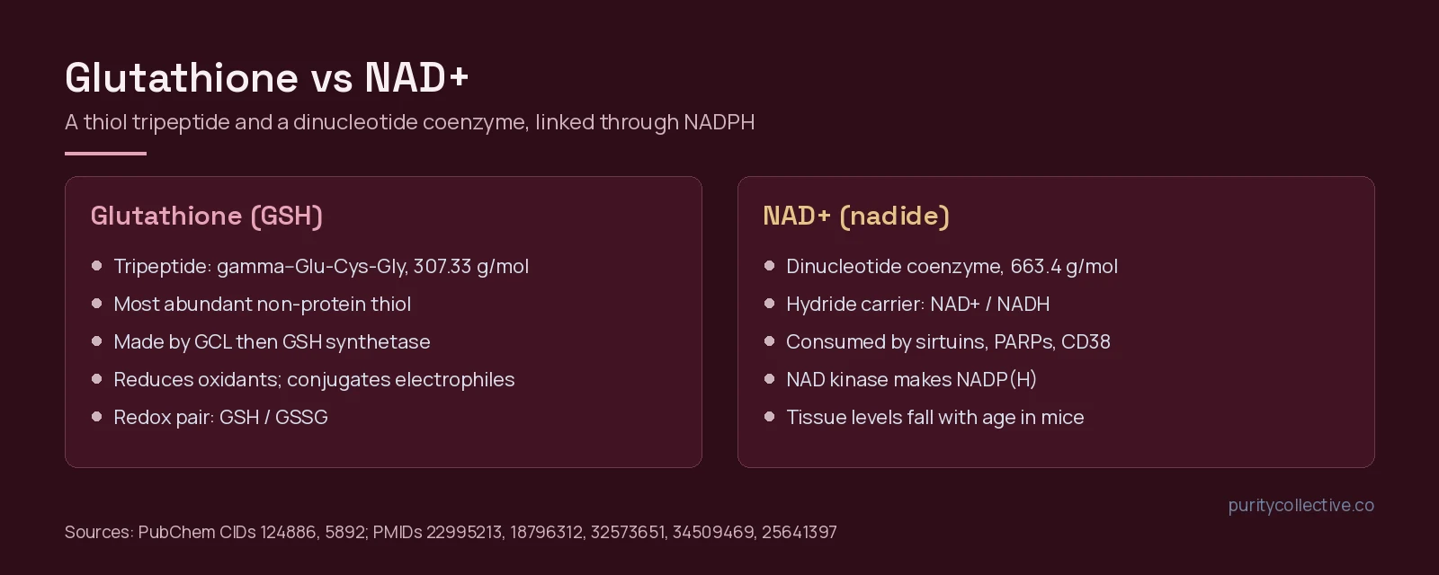
Glutathione vs NAD+ at a glance
| Feature | Glutathione (GSH) | NAD+ |
|---|---|---|
| Chemical class | Tripeptide thiol built through the intermediate γ-glutamylcysteine [1][10] | Dinucleotide coenzyme; pairs with its reduced form NADH [3] |
| Main role | Antioxidant defense, electrophile conjugation, redox signaling [1][2] | Redox coenzyme for glycolysis and mitochondrial respiration; co-substrate for signaling enzymes [3] |
| Redox pair | GSH and its disulfide GSSG [7][8] | NAD+/NADH; phosphorylated pool NADP+/NADPH [3][5] |
| Made by | Glutamate cysteine ligase (rate-limiting), then GSH synthetase; cysteine supply is a key limit [1] | De novo and salvage pathways; NAMPT is rate-limiting for salvage [4][9] |
| Consumed or modified by | Oxidants, conjugation reactions, protein S-glutathionylation [2][7] | Sirtuins, PARPs, SARM1 and CD38 [4] |
| Change with age (models) | Cellular GSH decline linked to GCL changes [10] | Modest tissue decline in aged mice with synthesis maintained [11] |
Identity of each compound
| Compound | Class | Formula | MW (g/mol) | PubChem CID |
|---|---|---|---|---|
| Glutathione (reduced, GSH) | Tripeptide thiol | C10H17N3O6S | 307.33 | 124886 |
| Glutathione disulfide (GSSG) | Oxidized dimer | C20H32N6O12S2 | 612.6 | 65359 |
| NAD+ (nadide) | Dinucleotide coenzyme | C21H27N7O14P2 | 663.4 | 5892 |
The GSSG row is there for a practical reason. Reduced glutathione oxidizes to its disulfide, and the two have very different masses, so a mass spectrometry trace on a glutathione batch should be read for both. NAD+ has its own pitfall: PubChem also lists a cationic record (CID 5893) at 664.4 g/mol, so the record named on a lab report should be matched to the form actually tested.
Glutathione: the cell’s thiol buffer
Glutathione is present in all mammalian tissues. A 2013 review lists its jobs as defense against oxidative stress, redox signaling, detoxification of xenobiotics, and roles in cell proliferation, apoptosis, immune function and fibrogenesis [1]. Synthesis happens in the cytosol in two enzyme steps. Glutamate cysteine ligase, built from a catalytic and a modifier subunit, makes γ-glutamylcysteine; glutathione synthetase completes the tripeptide. Cysteine availability and GCL activity set the pace, and Nrf2, AP-1 and NF-κB regulate the genes involved [1]. Our what is glutathione explainer covers the basics on their own.
Most of the action is in the thiol. GSH removes oxidants by reduction and electrophiles by conjugation [2], and it forms reversible mixed disulfides with protein cysteines, known as S-glutathionylation, which glutaredoxin enzymes add and remove as a regulatory switch [7]. Mitochondria import GSH made in the cytoplasm and use it to handle hydrogen peroxide from the respiratory chain; a long-term mismatch between mitochondrial ROS and mitochondrial GSH has been tied to apoptosis, necroptosis and ferroptosis in cell and animal work [8].
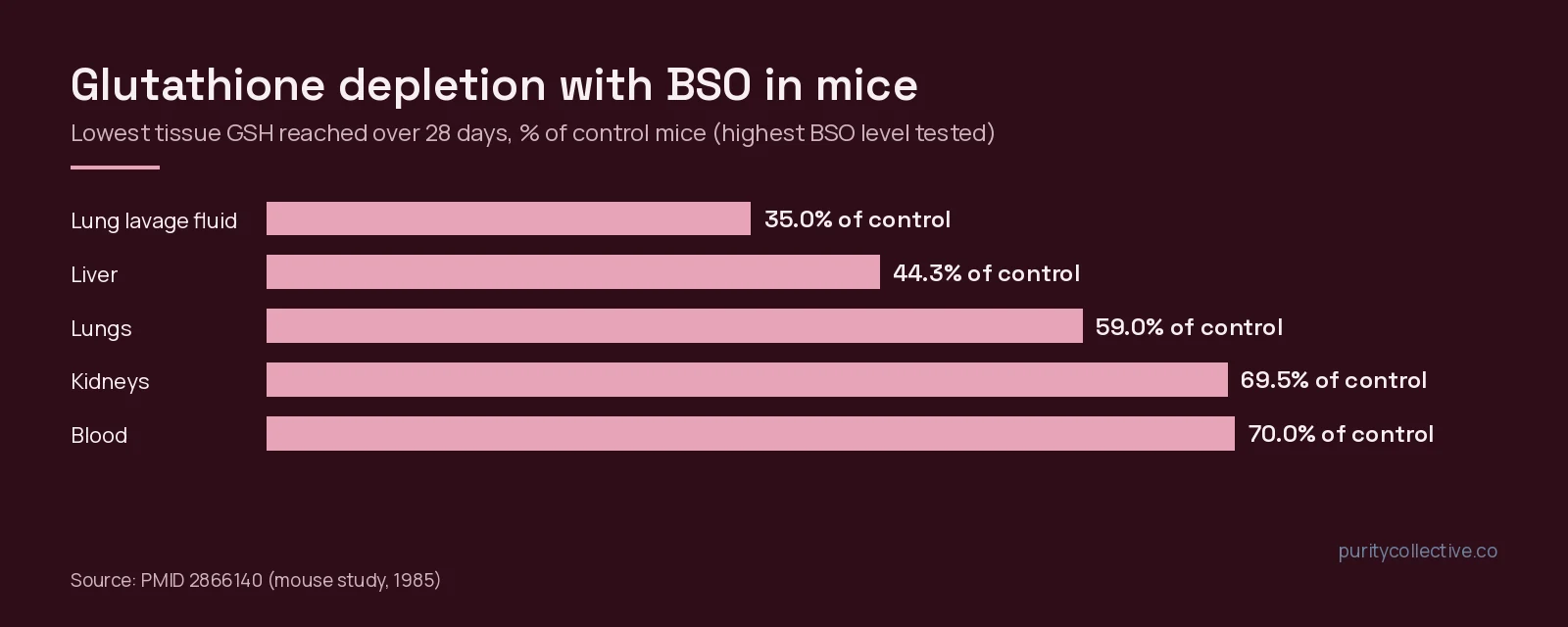
The classic tool for studying what happens without it is buthionine sulfoximine (BSO), which blocks GSH synthesis. In a 1985 mouse study, BSO in drinking water lowered GSH for up to 28 days, reaching 35% of control in lung lavage fluid and 44% in liver, and tissue levels returned to control after BSO was withdrawn [12].
NAD+: coenzyme and currency
NAD+ and NADH are coupled redox metabolites that drive oxidative, catabolic reactions in glycolysis and mitochondrial respiration. The same molecule is also a co-substrate: sirtuin deacetylases and PARP DNA-repair enzymes cleave it to nicotinamide as they work, so the cell must keep rebuilding it [3]. A 2021 review adds SARM1 and CD38 to the list of consumers and describes age-related NAD+ decline as an imbalance between biosynthesis and consumption, with lower NAMPT and more active PARPs and CD38 among the proposed causes [4]. Our NAD+ explainer goes further into the salvage pathway.
How large is the decline? In aged mice, isotope tracing found a median tissue NAD+ drop of about 30%. Circulating precursors and tryptophan-to-nicotinamide synthesis were unchanged, and in most tissues turnover of the smaller pool was faster, so absolute NAD+ synthesis was maintained. The authors read the drop as relatively subtle and driven by increased consumption [11]. Blocking one consumer has been tested directly: a specific CD38 inhibitor reversed tissue NAD+ decline and changed glucose tolerance, muscle and cardiac measures in naturally and prematurely aged mice, and those effects were lost when NAD+ synthesis was inhibited [13].
Where the two pools meet: NADPH
NAD+ and glutathione are linked by one enzyme step on each side. NAD kinase is the only enzyme that makes NADP, and because NADP cannot cross membranes, cells need compartment-specific NAD kinases, including a mitochondrial form identified from the human gene C5ORF33 [5]. The reduced form, NADPH, is the electron source glutathione reductase uses to reduce GSSG back to GSH, and NADPH pools in the cytosol and mitochondria are separate enough that a 2024 study built an organelle-specific assay to measure their consumption by that enzyme [6]. A 2008 enzymology review notes that cell redox status depends on both the absolute concentrations of these dinucleotides and their NAD/NADH and NADP/NADPH ratios [9].
For experimental design, this means an NAD+ manipulation can change glutathione recycling capacity downstream, while a glutathione manipulation, such as BSO depletion, does not directly change the NAD+ pool. Measuring GSH, GSSG and NADPH together helps separate the two.
What human studies have measured
| Study | Design | What was measured | Source |
|---|---|---|---|
| Oral glutathione | 6-month randomized, double-blind, placebo-controlled; 54 adults | GSH in whole blood, erythrocytes, plasma, lymphocytes and buccal cells | [14] |
| Nicotinamide riboside plus pterostilbene | 8-week randomized, double-blind, placebo-controlled; 120 adults aged 60–80 | Whole-blood NAD+ and safety | [15] |
| Three NAD+ precursors compared | 14-day randomized, open-label, placebo-controlled; 65 adults | Circulating NAD+; microbial metabolism ex vivo | [16] |
| Systematic review (2010–2025) | 33 human and 80 rodent intervention studies | Target engagement and functional outcomes of NAD-related compounds | [17] |
In the glutathione trial, blood GSH rose against baseline at both dose levels tested, mostly in a dose- and time-dependent way, and returned to baseline after a one-month washout [14]. In the precursor trials, whole-blood NAD+ rose dose-dependently with nicotinamide riboside plus pterostilbene [15], and in a three-way comparison nicotinamide riboside and nicotinamide mononucleotide raised circulating NAD+ while nicotinamide did not; ex vivo, both were converted by gut microbes to nicotinic acid [16]. These are biomarker results, not outcome results.
The 2026 systematic review is the useful reality check. Oral NAD+ precursors consistently showed biochemical target engagement in humans, but effects on functional, metabolic and vascular outcomes were heterogeneous and often null, and the authors found no eligible outcome trials of intravenous or intramuscular NAD+ itself [17]. Most of the NAD+ human literature is about precursors, not the dinucleotide sold as a research compound.
What the evidence does not show
- No study cited here compares glutathione with NAD+ directly; their link is biochemical, through NADPH [5][6].
- Age-related decline is clearer in models than its cause: the mouse tracing data point to consumption, not failed synthesis [11].
- Rodent results with NAD+ augmentation varied across models and endpoints [17].
- Blood glutathione and NAD+ levels are biomarkers; they do not establish an effect on any outcome [14][17].
For sourcing, the two need different checks. A glutathione research compound should be read for the GSH and GSSG forms, and an NAD+ research compound against the nadide record that matches the salt or form tested. When you buy research peptides online in the USA, ask for the batch-specific certificate rather than a generic sheet.
Frequently asked questions
Is glutathione a peptide?
Yes, in the narrow sense. Glutathione is a tripeptide of glutamate, cysteine and glycine, made inside cells by glutamate cysteine ligase and then glutathione synthetase (PMID 22995213). NAD+ is not a peptide; it is a dinucleotide coenzyme built from two nucleotides joined through their phosphate groups.
How are glutathione and NAD+ connected?
Through NADPH. NAD kinase phosphorylates NAD to NADP, and the reduced form NADPH supplies the electrons glutathione reductase uses to turn oxidized glutathione (GSSG) back into GSH (PMIDs 25641397, 39637235). The two pools are separate molecules but share one redox supply chain.
Do glutathione and NAD+ both decline with age?
Both have been reported to fall in aging models. Reviews describe a decline in cellular GSH linked to glutamate cysteine ligase, and lower tissue NAD+ in several organisms. In aged mice, isotope tracing found a median NAD+ drop of about 30% with production maintained and consumption increased (PMIDs 26845022, 34509469, 34559996).
What have human studies of these compounds measured?
Mostly blood biomarkers. A 6-month trial of oral glutathione measured GSH in blood and several cell types, and precursor trials measured whole-blood or circulating NAD+. A 2026 systematic review found no eligible outcome trials of intravenous or intramuscular NAD+ itself (PMIDs 24791752, 29184669, 41655607).
How should a lab confirm identity for each?
Check the batch certificate of analysis against PubChem: glutathione is CID 124886 (C10H17N3O6S, 307.33 g/mol) and NAD+ (nadide) is CID 5892 (C21H27N7O14P2, 663.4 g/mol). Purity Collective supplies both for laboratory research only, not for human or animal use.
References
- Lu SC (2013). Glutathione synthesis. Biochim Biophys Acta. PMID: 22995213. doi: 10.1016/j.bbagen.2012.09.008.
- Forman HJ, Zhang H, Rinna A (2009). Glutathione: overview of its protective roles, measurement, and biosynthesis. Mol Aspects Med. PMID: 18796312. doi: 10.1016/j.mam.2008.08.006.
- Griffiths HBS, Williams C, King SJ et al. (2020). Nicotinamide adenine dinucleotide (NAD+): essential redox metabolite, co-substrate and an anti-cancer and anti-ageing therapeutic target. Biochem Soc Trans. PMID: 32573651. doi: 10.1042/BST20190033.
- Strømland Ø, Diab J, Ferrario E et al. (2021). The balance between NAD(+) biosynthesis and consumption in ageing. Mech Ageing Dev. PMID: 34509469. doi: 10.1016/j.mad.2021.111569.
- Zhang R (2015). MNADK, a Long-Awaited Human Mitochondrion-Localized NAD Kinase. J Cell Physiol. PMID: 25641397. doi: 10.1002/jcp.24926.
- Ting KKY, Floro E, Dow R et al. (2024). Measuring the rate of NADPH consumption by glutathione reductase in the cytosol and mitochondria. PLoS One. PMID: 39637235. doi: 10.1371/journal.pone.0309886.
- Chai YC, Mieyal JJ (2023). Glutathione and Glutaredoxin-Key Players in Cellular Redox Homeostasis and Signaling. Antioxidants (Basel). PMID: 37627548. doi: 10.3390/antiox12081553.
- Chen TH, Wang HC, Chang CJ et al. (2024). Mitochondrial Glutathione in Cellular Redox Homeostasis and Disease Manifestation. Int J Mol Sci. PMID: 38279310. doi: 10.3390/ijms25021314.
- Magni G, Orsomando G, Raffelli N et al. (2008). Enzymology of mammalian NAD metabolism in health and disease. Front Biosci. PMID: 18508649. doi: 10.2741/3143.
- Ferguson G, Bridge W (2016). Glutamate cysteine ligase and the age-related decline in cellular glutathione: The therapeutic potential of γ-glutamylcysteine. Arch Biochem Biophys. PMID: 26845022. doi: 10.1016/j.abb.2016.01.017.
- McReynolds MR, Chellappa K, Chiles E et al. (2021). NAD(+) flux is maintained in aged mice despite lower tissue concentrations. Cell Syst. PMID: 34559996. doi: 10.1016/j.cels.2021.09.001.
- Sun JD, Ragsdale SS, Benson JM et al. (1985). Effects of the long-term depletion of reduced glutathione in mice administered L-buthionine-S,R-sulfoximine. Fundam Appl Toxicol. PMID: 2866140. doi: 10.1016/0272-0590(85)90173-3.
- Tarragó MG, Chini CCS, Kanamori KS et al. (2018). A Potent and Specific CD38 Inhibitor Ameliorates Age-Related Metabolic Dysfunction by Reversing Tissue NAD(+) Decline. Cell Metab. PMID: 29719225. doi: 10.1016/j.cmet.2018.03.016.
- Richie JP Jr, Nichenametla S, Neidig W et al. (2015). Randomized controlled trial of oral glutathione supplementation on body stores of glutathione. Eur J Nutr. PMID: 24791752. doi: 10.1007/s00394-014-0706-z.
- Dellinger RW, Santos SR, Morris M et al. (2017). Repeat dose NRPT (nicotinamide riboside and pterostilbene) increases NAD(+) levels in humans safely and sustainably: a randomized, double-blind, placebo-controlled study. NPJ Aging Mech Dis. PMID: 29184669. doi: 10.1038/s41514-017-0016-9.
- Christen S, Redeuil K, Goulet L et al. (2026). The differential impact of three different NAD(+) boosters on circulatory NAD and microbial metabolism in humans. Nat Metab. PMID: 41540253. doi: 10.1038/s42255-025-01421-8.
- Gallagher C, Emmanuel OO (2026). NAD⁺ supplementation for anti-aging and wellness: A PRISMA-guided systematic review of preclinical and clinical evidence. Ageing Res Rev. PMID: 41655607. doi: 10.1016/j.arr.2026.103057.