
For laboratory research only. Not for human or animal use. This article gives no dosing, administration or therapeutic advice.
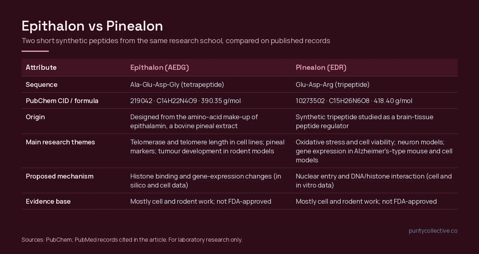
Short answer: Epithalon and Pinealon are two different short synthetic peptides. Epithalon is a four-amino-acid chain (Ala-Glu-Asp-Gly, often written AEDG); Pinealon is a three-amino-acid chain (Glu-Asp-Arg, or EDR). They share a research lineage, not a structure. Epithalon’s literature is mostly about telomerase, pineal-gland markers and tumour development in rodents. Pinealon’s literature is mostly about oxidative stress and nerve-cell models. Almost all of it is cell and animal work, and most of it comes from one research group.
Two peptides, one research programme
Both peptides come out of the “peptide bioregulator” programme developed in St Petersburg, Russia. Epithalon was synthesised to match the amino-acid composition of epithalamin, a polypeptide extract of bovine pineal gland[1]. A 2025 review in the International Journal of Molecular Sciences summarises 25 years of in vitro, in vivo and in silico work on it, and notes that physico-chemical and structural studies of the peptide remain limited[1].
Pinealon is a synthetic tripeptide. Despite the name, its published work focuses on brain cells rather than the pineal gland: cerebellar granule cells, PC12 cells and cortical cultures[2][3]. A 2020 review frames EDR as a peptide that may enter cells and bind histones or nucleic acids, which its authors link to changes in gene expression[4].
So the useful framing for a lab is not “which one is better” but “which question does each one’s literature actually address”.
Structure and identity
| Attribute | Epithalon | Pinealon |
|---|---|---|
| Sequence | Ala-Glu-Asp-Gly (AEDG) | Glu-Asp-Arg (EDR) |
| Length | 4 residues | 3 residues |
| PubChem CID | 219042 | 10273502 |
| Molecular formula | C14H22N4O9 | C15H26N6O8 |
| Molecular weight (PubChem) | 390.35 g/mol | 418.40 g/mol |
| Charge character | Two acidic residues (Glu, Asp) | Two acidic residues plus one basic residue (Arg) |
| Also written as | Epitalon, Epithalone, AEDG peptide | EDR peptide, Glu-Asp-Arg |
Because the two sequences are so short, a mass-spectrometry identity result is the quickest way to tell them apart on paper: the expected masses differ by about 28 g/mol. Our guide on how to read a peptide COA explains where to find the identity and purity results on a certificate.
What the Epithalon literature examines
Telomerase and telomere length in cell culture
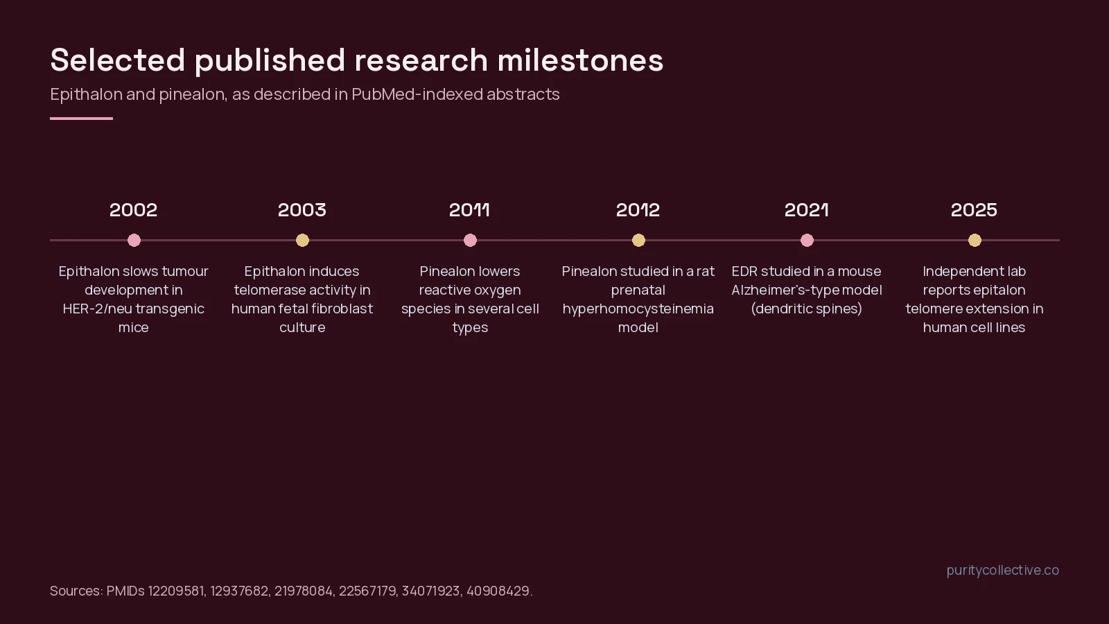
The most cited Epithalon finding is a 2003 report that adding the peptide to telomerase-negative human fetal fibroblast cultures induced expression of the telomerase catalytic subunit, telomerase activity and telomere elongation[5]. That was a single cell-culture study from the originating group.
A 2025 paper in Biogerontology, from a separate group, revisited the question in breast cancer cell lines (21NT, BT474) and in normal epithelial and fibroblast cells. It reported dose-dependent telomere extension in the normal cells through hTERT and telomerase upregulation, and telomere extension in the cancer lines through a different route, alternative lengthening of telomeres (ALT)[6]. That cancer-cell finding is a reason for caution as much as interest: telomere maintenance is a hallmark of tumour cells, so the result does not read as simply favourable.
Gene expression and stem-cell models
In human gingival mesenchymal stem cells, AEDG increased mRNA and protein for neuronal differentiation markers (Nestin, GAP43, β-tubulin III, doublecortin), and molecular modelling suggested binding to linker histones H1/3 and H1/6[7]. In long-term cultures of oral stem cells at passage 25, AEDG lowered expression of the senescence markers p16 and p21 compared with untreated controls[8]. Both are in vitro studies, and the histone-binding model is computational.
Pineal-gland markers
In rats, intranasal Epithalon produced a small but significant rise in the C-Fos protein in pinealocytes only in animals under osmotic stress, not in unstressed animals[9]. In organotypic pineal cultures from 3-month-old rats, AEDG increased the proliferation marker Ki-67 and was the only peptide tested that increased CGRP synthesis; Pinealon (EDR), tested in the same study, did not show the CGRP effect[10]. That paper is one of the few direct, same-study comparisons of the two peptides.
Rodent tumour and life-span studies
In HER-2/neu transgenic mice, Epithalon reduced the cumulative number and maximum size of spontaneous mammary tumours compared with saline[11]. In female C3H/He mice, fewer treated animals had malignant tumours, and no metastases were seen in the treated group[12]. The results are not uniform, though: in female rats kept under a standard light-dark cycle, Epithalon did not change life span and had virtually no effect on spontaneous tumours; effects were reported only in rats kept under natural northern or constant light[13]. These studies used small groups and come from the same research network.
Oocyte and retinal-cell models
Two newer in vitro studies from other groups examined oxidative stress. In aging mouse oocytes, Epitalon in the culture medium lowered reactive oxygen species and reduced spindle defects over 24 hours[14]. In a high-glucose model using a human retinal pigment epithelial cell line (ARPE-19), it restored delayed wound closure and reduced markers of epithelial-mesenchymal transition[15]. The authors of the second study note that more mechanistic and safety work is needed[15].
What the Pinealon literature examines
Oxidative stress and cell viability
The founding Pinealon paper reported dose-dependent limits on reactive oxygen species build-up in rat cerebellar granule cells, neutrophils and PC12 cells under oxidative stress, alongside less necrotic cell death and delayed ERK1/2 activation[2]. The authors noted that the ROS effect plateaued at lower concentrations while cell-cycle changes continued at higher ones, and proposed a direct interaction with the genome[2].
Animal models
In a rat model of prenatal hyperhomocysteinemia (pregnant rats given a methionine-loaded diet), offspring from the Pinealon-treated group showed better spatial orientation and learning, and their isolated cerebellar neurons accumulated less ROS[16]. In the 5xFAD mouse model of Alzheimer’s-type pathology, EDR and a related tripeptide (KED) were reported to prevent dendritic-spine loss, and docking studies placed EDR binding sites in promoter regions of genes such as CASP3, APOE and SOD2[17].
Cortical cells and induced neurons
EDR stimulated serotonin expression in aging cortical cell cultures, with docking pointing at the tryptophan hydroxylase gene[3]. In a 2024 model of neurons transdifferentiated from the skin fibroblasts of elderly donors, EDR reduced oxidative DNA damage, and all three peptides tested (EDR, KED and AEDG) increased dendritic branching. The same study reported that the tripeptides had no effect on mitochondrial or lysosomal activity or on p16 levels in those neurons[18], a null result worth keeping in view.
Shared mechanism claims, and their limits
Both peptides are proposed to work by entering the cell nucleus and interacting with DNA or histones. One fluorescence study showed that labelled Epithalon and Pinealon both entered the cytoplasm, nucleus and nucleolus of HeLa cells, and that each bound preferentially to certain DNA sequences in vitro[19]. That is evidence of cell entry and DNA interaction in a test system; it does not show that the gene-expression changes reported elsewhere follow from that binding. Much of the gene-target work for both peptides relies on molecular docking[7][17][3].
Side-by-side: how the evidence compares
| Question | Epithalon (AEDG) | Pinealon (EDR) |
|---|---|---|
| Main model types | Human and rodent cell lines; mice and rats | Rat neurons, PC12 cells; rats; 5xFAD mice; induced human neurons |
| Headline research theme | Telomerase and telomere length[5][6] | Oxidative stress in neural cells[2] |
| Independent groups | Some (2022, 2025 cell studies)[14][6] | Few; mostly the originating group[4] |
| Mixed or null results | No life-span change under standard lighting in rats[13]; ALT activation in cancer lines[6] | No effect on mitochondria, lysosomes or p16 in induced neurons[18]; no CGRP effect in pineal culture[10] |
| Regulatory status | Not FDA-approved | Not FDA-approved |
Choosing between them for a study design
- Telomere, telomerase or senescence-marker work in cell culture: Epithalon has the larger and more varied record, including one independent 2025 cell-line study[6].
- Oxidative stress in neural cell models: Pinealon’s literature sits closer to that question[2][18].
- Direct comparisons: few exist. The pineal organotypic culture study is one, and it found different marker profiles for the two peptides[10].
Whichever one a lab picks, design controls with the evidence gaps in mind: small published sample sizes, limited replication outside one research school, and heavy reliance on docking for mechanism. Read more on research-grade sourcing in what research peptides are.
Documentation to check
For either peptide, match the sequence and molecular weight on the certificate to PubChem, check that the mass-spectrometry result fits the expected mass, read the HPLC purity figure, and confirm that the batch number on the vial is the one on the report. Purity Collective lists Epithalon 10mg, Epithalon 50mg and Pinealon 10mg for laboratory research. You can buy research peptides online in the USA from our catalogue with batch documentation.
Frequently asked questions
What is the difference between Epithalon and Pinealon?
Epithalon is the tetrapeptide Ala-Glu-Asp-Gly (AEDG) and Pinealon is the tripeptide Glu-Asp-Arg (EDR). They share no sequence. Published work on Epithalon centres on telomerase, pineal markers and rodent tumour models, while Pinealon work centres on oxidative stress and neuron models.
Are Epithalon and Pinealon the same peptide?
No. They are different molecules with different PubChem records (CID 219042 for Epithalon, CID 10273502 for Pinealon). The names are sometimes confused because both come from the same short-peptide research programme in St Petersburg and both are linked to the pineal gland in their names.
Has either peptide been approved by the FDA?
No. Neither Epithalon nor Pinealon is an FDA-approved drug. Most published data come from cell cultures and rodent models, and both are sold by Purity Collective strictly for laboratory research.
Who has published research on these peptides?
Most papers come from one research school led by Vladimir Khavinson and colleagues, sometimes with Italian collaborators. A 2025 Biogerontology paper from a separate group reported telomere-length changes with Epithalon in human cell lines. Independent replication is still limited for both peptides.
What should a lab check before using either peptide in a study?
Confirm the sequence and molecular weight against PubChem, read the HPLC purity and mass-spectrometry identity results on the certificate of analysis, and make sure the batch number on the vial matches the report.
References
- Araj SK, Brzezik J, Mądra-Gackowska K et al. (2025). Overview of Epitalon-Highly Bioactive Pineal Tetrapeptide with Promising Properties. Int J Mol Sci. PMID: 40141333. doi: 10.3390/ijms26062691.
- Khavinson V, Ribakova Y, Kulebiakin K et al. (2011). Pinealon increases cell viability by suppression of free radical levels and activating proliferative processes. Rejuvenation Res. PMID: 21978084. doi: 10.1089/rej.2011.1172.
- Khavinson VKh, Lin’kova NS, Tarnovskaya SI et al. (2014). Short peptides stimulate serotonin expression in cells of brain cortex. Bull Exp Biol Med. PMID: 24909721. doi: 10.1007/s10517-014-2496-y.
- Khavinson V, Linkova N, Kozhevnikova E et al. (2020). EDR Peptide: Possible Mechanism of Gene Expression and Protein Synthesis Regulation Involved in the Pathogenesis of Alzheimer’s Disease. Molecules. PMID: 33396470. doi: 10.3390/molecules26010159.
- Khavinson VKh, Bondarev IE, Butyugov AA (2003). Epithalon peptide induces telomerase activity and telomere elongation in human somatic cells. Bull Exp Biol Med. PMID: 12937682. doi: 10.1023/a:1025493705728.
- Al-Dulaimi S, Thomas R, Matta S et al. (2025). Epitalon increases telomere length in human cell lines through telomerase upregulation or ALT activity. Biogerontology. PMID: 40908429. doi: 10.1007/s10522-025-10315-x.
- Khavinson V, Diomede F, Mironova E et al. (2020). AEDG Peptide (Epitalon) Stimulates Gene Expression and Protein Synthesis during Neurogenesis: Possible Epigenetic Mechanism. Molecules. PMID: 32019204. doi: 10.3390/molecules25030609.
- Sinjari B, Diomede F, Khavinson V et al. (2020). Short Peptides Protect Oral Stem Cells from Ageing. Stem Cell Rev Rep. PMID: 31677028. doi: 10.1007/s12015-019-09921-3.
- Sibarov DA, Kovalenko RI, Malinin VV et al. (2002). Epitalon influences pineal secretion in stress-exposed rats in the daytime. Neuro Endocrinol Lett. PMID: 12500171.
- Khavinson VKh, Linkova NS, Chalisova NI et al. (2011). Effect of short peptides on expression of signaling molecules in organotypic pineal cell culture. Bull Exp Biol Med. PMID: 22803060. doi: 10.1007/s10517-011-1473-y.
- Anisimov VN, Khavinson VK, Provinciali M et al. (2002). Inhibitory effect of the peptide epitalon on the development of spontaneous mammary tumors in HER-2/neu transgenic mice. Int J Cancer. PMID: 12209581. doi: 10.1002/ijc.10570.
- Kossoy G, Anisimov VN, Ben-Hur H et al. (2006). Effect of the synthetic pineal peptide epitalon on spontaneous carcinogenesis in female C3H/He mice. In Vivo. PMID: 16634527.
- Vinogradova IA, Bukalev AV, Zabezhinski MA et al. (2007). Effect of Ala-Glu-Asp-Gly peptide on life span and development of spontaneous tumors in female rats exposed to different illumination regimes. Bull Exp Biol Med. PMID: 18856211. doi: 10.1007/s10517-007-0441-z.
- Yue X, Liu SL, Guo JN et al. (2022). Epitalon protects against post-ovulatory aging-related damage of mouse oocytes in vitro. Aging (Albany NY). PMID: 35413689. doi: 10.18632/aging.204007.
- Gatta M, Dovizio M, Milillo C et al. (2025). The Antioxidant Tetrapeptide Epitalon Enhances Delayed Wound Healing in an in Vitro Model of Diabetic Retinopathy. Stem Cell Rev Rep. PMID: 40493162. doi: 10.1007/s12015-025-10911-x.
- Arutjunyan A, Kozina L, Stvolinskiy S et al. (2012). Pinealon protects the rat offspring from prenatal hyperhomocysteinemia. Int J Clin Exp Med. PMID: 22567179.
- Khavinson V, Ilina A, Kraskovskaya N et al. (2021). Neuroprotective Effects of Tripeptides-Epigenetic Regulators in Mouse Model of Alzheimer’s Disease. Pharmaceuticals (Basel). PMID: 34071923. doi: 10.3390/ph14060515.
- Kraskovskaya N, Linkova N, Sakhenberg E et al. (2024). Short Peptides Protect Fibroblast-Derived Induced Neurons from Age-Related Changes. Int J Mol Sci. PMID: 39518916. doi: 10.3390/ijms252111363.
- Fedoreyeva LI, Kireev II, Khavinson VKh et al. (2011). Penetration of short fluorescence-labeled peptides into the nucleus in HeLa cells and in vitro specific interaction of the peptides with deoxyribooligonucleotides and DNA. Biochemistry (Mosc). PMID: 22117547. doi: 10.1134/S0006297911110022.